20 Jan What is OCT? | Optical Coherence Tomography
OCT stands for optical coherence tomography and is an important diagnostic tool used by most eye doctors, but is exceptionally useful to the retina specialist.
OCT can be used alone to study the retina or in conjunction with a variety of other testing modalities to include retinal digital photography and fluorescein angiography. This non-invasive test may be used to help diagnose and treat glaucoma and retinal diseases although the testing procedure is not exactly the same.
The test provides information in “high-definition” due to its ability to examine and provide information down to the micrometer level. Before OCT, this resolution of testing was not possible.
Optical coherence tomography may be performed on the retina and separately focused on the optic nerve (glaucoma).
This article focuses on OCT for retinal diseases and how it has become the mainstay of diagnostic testing which offers different information than a fluorescein angiogram.
What OCT
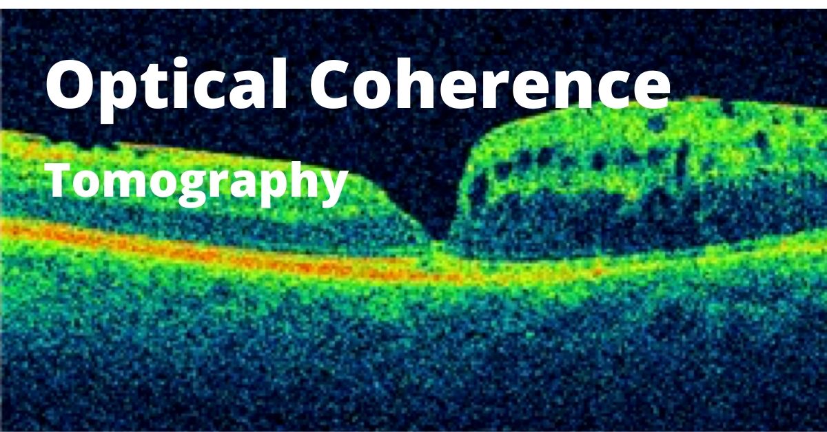
This is a painless non-invasive test using light waves to examine the different layers of the retina. The surface topography can also be examined.
By the way, tomography allows us to examine a tissue in cross-section, whereas topography allows us to examine the surface of a tissue.
Results of an OCT are best if the pupils are dilated, but dilation is not necessary.
An OCT is obtained by placing resting your chin on the machine while keeping your eyes and head as still as possible. Nothing will touch your eyes.
The scan will take several minutes while you look at a target keeping your eyes still.
When to Use OCT
OCT is very useful to the retina specialist to evaluate common retinal diseases such as:
- Macular holes
- Macular pucker
- Macular edema caused by various conditions such as retinal vascular occlusions (RVO)
- Macular degeneration (ARMD)
- Central serous retinopathy (CSR)
- Diabetic retinopathy
- Vitreomacular traction (VMT)
Optical coherence tomography is extremely useful in monitoring the effectiveness of a treatment and answers the question, is the patient getting better?
OCT has a few limitations. The test is very difficult to perform in situations where light is blocked from entering the eye, such as: dense cataracts or vitreous hemorrhage.
No Comments