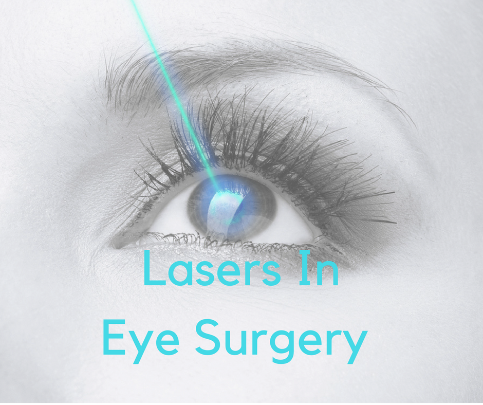
Categories
Cataract Cataract Surgery Surgery Treatments Vision Correction

Lasers Used in Ophthalmology

Lasers Used in Ophthalmology

Lasers used in ophthalmologyLaser eye surgery can mean many more things than laser vision correction.  Lasers are used in almost all parts of the eye to treat eye disease such as glaucoma, refractive eye surgery and many diseases of the retina.

The following is a short review discussing the many different lasers and how we use them in ophthalmology.

Laser Vision Correction

Laser vision correction is a very common laser eye surgery.  PRK and LASIK are often advertised to reduce a patient’s need and dependance upon glasses (or contact lenses).

Both the femtosecond and excimer lasers are used to perform laser vision correction.  The lasers reshape the corneal curvature which heavily influences the refraction of your eye.

Laser vision correction reduces the need for glasses in patients who have nearsightedness (myopia), farsightedness (hyperopia) and astigmatism.  Certain corneal scars can also be removed with the laser.

Laser Cataract Surgery

Modern cataract surgery now involve the accuracy and dependability of the femtosecond laser.  The laser can be used in crucial parts of the cataract removal process:

  • creating a corneal incision (the first step)
  • creating the capsulorhexis (the opening of the lens capsule)
  • liquefying the cataract
  • treating astigmatism, if needed

The cataract can be compared to an M & M candy.  A candy coating surrounding a chocolate center.  In this analogy, the cataract is enclosed by the lens capsule:  the cataract is the chocolate center and the lens capsule is the outside candy coating.  During cataract surgery, the “capsulorhexis” is the opening created in the front side of the cataract, the candy coating, which allow access to the cataract (chocolate) held within.  Once the chocolate is removed, the IOL is placed within the candy coating, lens capsule, keeping the IOL in place.

After Cataract – Posterior Capsular Opacification

During cataract surgery, the intraocular implant is placed inside a clear tissue called the lens capsule.  Just like “Saran Wrap,” this tissue is crystal clear.  With time, the tissue often becomes cloudy and blurs the vision.  This “after cataract” mimics the same symptoms of the original cataract, but vision can be restored with Nd:YAG laser which clears the cloudy lens capsule.

Glaucoma

Several different lasers can be used to treat different types of glaucoma.

Open angle glaucoma, the most common form of glaucoma in the United States, is usually treated with eye drops to lower the eye pressure.  When the eye drops fail to work, ALT (Argon Laser Trabeculoplasty), SLT (Selective Laser Trabeculoplasty) or MLT (Minipulse Laser Trabeculoplasty) can be used to treat patients with open angle glaucoma.

These lasers treat the drainage part of the eye, called the trabecular meshwork, to increase the absorption of internal fluid (aqueous humor) and decrease the pressure of the eye.

The laser procedures are similar and differ by the types of laser used:  Argon or diode laser, Nd:YAG laser or the minipulse laser.

Laser can also be used to reduce the production of aqueous humor by treating the ciliary body.  The ciliary body is located behind the iris and can be treated with argon or diode laser to reduce the production of fluid, thereby reducing eye pressure.  This form of treatment is often done in conjunction with cataract surgery.

Narrow angle glaucoma is common in asians and patients with hyperopia.  In this scenario, fluid is blocked from migrating through the pupil to the anterior chamber to

Retina and Vitreous

The argon laser, diode laser and minipulse laser are used to treat patients with diabetic retinopathy, swelling from various retinal vascular disorders and others.  Tears in the retina are usually treated with the argon or diode laser.

Nd:YAG vitreolysis is performed for certain opacities within the vitreous.


[siteorigin_widget class=”WP_Widget_Media_Image”][/siteorigin_widget]
Categories
“Other” Eye Conditions Vision Correction

First Laser Vision Correction Performed 30 Years Ago

Laser Vision Correction Started 30 Years AgoThe first laser vision correction patient had PRK performed on one eye 30 years ago on March 25, 1988.  Vision correction procedures have been performed since the mid 1950s, but never before using a laser.

PRK (photorefractive keratectomy) is still performed today though LASIK has become the modern favorite, at least in the United States.

Innovative Idea

The idea to use a laser on human tissue began in 1983 with the publishing of a scientific paper proposing the idea.  The excimer laser had been already developed, but was used for industrial purposes to manufacture microelectronic devices and semi-conductors.

What if the precision of the excimer laser could be used to reshape the cornea, and hence, change the refractive power of the eye?

The team of researchers practiced and practiced, first on plastic blocks then on cadaver animal and human eyes.  Finally, the “procedure” was ready for live human testing.

How would they go about getting permission?

Mrs. Cassady Had Cancer

The first patient, Mrs. Cassady, to have laser vision correction had cancer of the eye socket.  Treatment for the cancer behind her eye was to include removal of the healthy eye.

Mrs. Cassady gave her permission to have PRK performed to her eye before the eye was to be removed.  The FDA agreed.

The laser surgery was successful and paved the way for eventual clinical trials for the excimer to be used clinically.

All thanks to the bravery of Mrs. Cassady.

PRK Develops into LASIK

PRK and LASIK (laser-assisted in situ keratomileusis) are very similar.  Both use the excimer laser to reshape the cornea to accurately change the refractive power of the eye.

The curvature of the cornea, along with a healthy tear film, is responsible for about 75% of the total focusing power of the eye.  Changes in the curvature are responsible for the “correction” using the laser.

While the visual results are essentially identical, LASIK requires formation of a corneal flap, whereas PRK does not.  The hinged flap, like a man hole cover is moved out of the way during the laser reshaping and then replaced.  In PRK the surface tissue is ablated and not replaced.  The corneal surface will heal by itself.

The corneal flap:

  • Allows for faster healing
  • Improves vision faster
  • Causes less discomfort

On the other hand…not all patients can have LASIK.   Patients who are considering LASIK must have a thick enough cornea for a flap to be created, that is, some patients with insufficient corneal thickness or too high a prescription can only consider PRK.

Randall Wong, M.D.
Retina Specialist
Fairfax, Virginia, USA

Verified by MonsterInsights