07 Jan Blood in the Retina: You Make the Call
A patient of mine returned this morning with complaints of decreased vision in the right eye. She is 84 years old, has a history of smoking and noted some “blurriness” in the right eye for the past few months. With both eyes open, however, she sees pretty well.
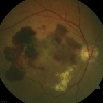
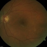
The first thing we did was examine her. This is a retinal photograph of the right eye. What do you see? There is blood distributed underneath the retina. The white areas are signs of chronic leakage and are called exudates. The left eye, pictured below is normal. What do we need to do next?
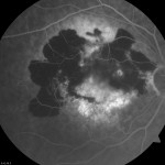
The next step is to perform a fluorescein angiogram. The retinal hemorrhage is characteristic of bleeding due to “wet” macular degeneration. The fluorescein angiogram should show evidence of neovascularization and provide evidence of the correct wet macular degeneration diagnosis.
What Does This Mean? The first picture is typical (although exaggerated) of patients with wet macular degeneration. While most do not bleed, the blood doesn’t cause any further permanent damage. The fluorescein angiogram is the best test to confirm “neovascularization” in the retina, the source of bleeding. Years ago (say around 2000) there would be nothing to do. Laser photocoagulation would not be helpful as there is too much blood. Now, the best choice is to consider intravitreal injections of Avastin (anti-VEGF).
I’d recommend an initial course of three injections of Avastin. We’ll be starting within the next week.
“Randy”
Randall V. Wong, M.D.
Ophthalmologist, Retina Specialist
Fairfax, Virginia


![Reblog this post [with Zemanta]](http://img.zemanta.com/reblog_e.png?x-id=588e60a9-d458-4a7c-af38-facfb5894fec)
No Comments